


Sponsored by:

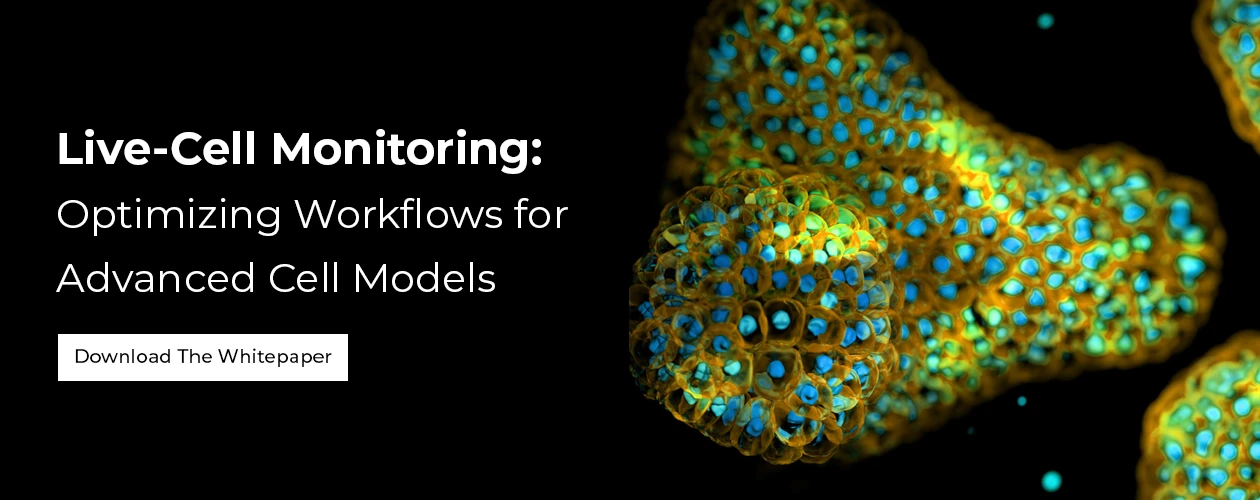
Live-Cell Monitoring: Optimizing Workflows for Advanced Cell Models
In this in-depth white paper from Sartorius, you will discover how live-cell monitoring and real-time imaging can optimize advanced cell culture workflows—without disturbing your samples.
Advanced cell models such as organoids, spheroids, stem cells, and iPSC-derived systems are transforming biomedical research, drug discovery, and regenerative medicine. However, as these models become more complex, ensuring consistent quality control (QC) and capturing dynamic cellular events throughout long workflows has never been more critical.
In this in-depth white paper from Sartorius, you will discover how live-cell monitoring and real-time imaging can optimize advanced cell culture workflows—without disturbing your samples. By continuously visualizing cellular health, morphology, differentiation, activation, and phenotypic changes, researchers can make better-informed decisions, reduce assay failure, and improve data reliability.
What You’ll Learn in This White Paper:
- Why live-cell monitoring is essential for QC across cell culture, manipulation, and assay stages
- How to capture dynamic cellular events during differentiation, activation, and reprogramming
- Best practices for monitoring iPSC generation, stem cell differentiation, and rare cellular events
- Strategies for analyzing and standardizing 3D models, spheroids, and organoids
- How label-free and fluorescent live-cell imaging enhances reproducibility and confidence in results